What is Spondylolysis?
19/02/2024
What is Spondylolysis?
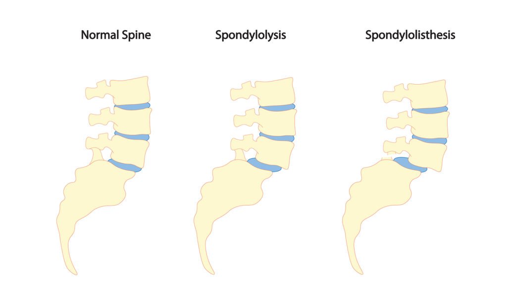
Spondylolysis, which is a stress fracture of the spine due to overuse, is one of the most common causes of back pain in adolescent athletes. The stress fracture is in the posterior part of the spine, in an area called the pars interarticularis. In almost all cases, it occurs at the L4 or L5 vertebrae, which are the lowest parts of the lumbar spine.

Approximately 80% of the time, the injury involves both the left and right pars interarticularis at the given level, which can lead to forward slippage of the vertebrae; if this occurs, the injury is called spondylolisthesis. The severity of spondylolisthesis is graded I to V depending on how far the vertebra has slipped anteriorly.
Why does it occur?
Spondylolysis and spondylolisthesis are caused by repetitive hyperextension and twisting of the spine. So, it is often seen in athletes that participate in sports such as gymnastics, figure skating, ballet and other dance, wrestling, diving, and football. When an adolescent is growing, the pelvis tilts anteriorly as the lordosis in the lumbar spine increases. Lordosis is what causes the lower back to look “swayed” forwards; it is normal to have some lordosis in the lumbar spine, but can cause problems when there is excessive lordosis. This change causes more compression in the posterior spine, which can increase the risk of stress fracture in this area. Additionally, during the growth spurt, bone grows faster than the muscles and tendons which leads to some muscular imbalances, including tightness in the hip flexors and weakness of the abdominals and gluteal muscles. Another risk factor for developing spondylolysis or spondylolisthesis is having Scheuermann’s kyphosis in the thoracic (upper) spine, as the lumbar spine may develop excess lordosis in order to compensate and “straighten” the spine.
How is it diagnosed?
Symptoms are usually back pain with extension of the spine that worsens with activity and pain in the middle of the back that sometimes radiates to the buttocks or down one leg. A test called the Stork test where a patient stands on one leg and extends the spine can be used in the diagnosis process. There is usually not pain with spinal flexion (bending forwards).
Imaging is often used to confirm the diagnosis if the pain lasts more than a few weeks. The most sensitive type of imaging to detect a spondylolysis is a SPECT scan, or single-photon emission computed tomography; however, these do expose the patient to a significant amount of radiation. Therefore, MRI (magnetic resonance imaging) is commonly used to diagnose spondylolysis as it can help with diagnosis without radiation exposure.
What is the treatment?
Most treatment plans consist of activity modification and physiotherapy, with braces sometimes being used for additional healing support. Physiotherapy includes strengthening of the core, spinal, and hip musculature and improving flexibility of any muscles that are tight. Activity modification often involves being out of sports for a period of time, or at least avoiding any activities that cause pain. The physiotherapist will help with activity modification in the early stages of recovery as well as helping to assist with body mechanics specific to the patient’s sport or lifestyle to decrease risk of the pain returning.

References for text:
D'Hemecourt PA, Gould LE, Bottin NM. Spondylolysis. In: Stein CJ, Stracciolini A, Ackerman KE, eds. The Young Female Athlete, Contemporary Pediatric and Adolescent Sports Medicine. Switzerland: Springer; 2016: 183-206.
Spondylolysis and Spondylolisthesis. American Academy of Orthopaedic Surgeons. Updated August 2020. https://orthoinfo.aaos.org/en/diseases--conditions/spondylolysis-and-spondylolisthesis/
References for Photo:
Spondylolysis and Spondylolisthesis. American Academy of Orthopaedic Surgeons. Updated August 2020. https://orthoinfo.aaos.org/en/diseases--conditions/spondylolysis-and-spondylolisthesis/
